
FreeLesson 1 of 36 min read
The main parts of an animal cell and what they do
Meet the nucleus, membrane, cytoplasm, mitochondria and ribosomes, and see how they work together to keep a cell alive.
Every living thing is made of cells, and every life process happens inside them. Moving, growing, healing and digesting all depend on them. Once you know what each part of a cell does, the rest of biology makes far more sense. Why do muscles tire? Why do we need oxygen? The answers start here.
A diagram is a model, not a photograph
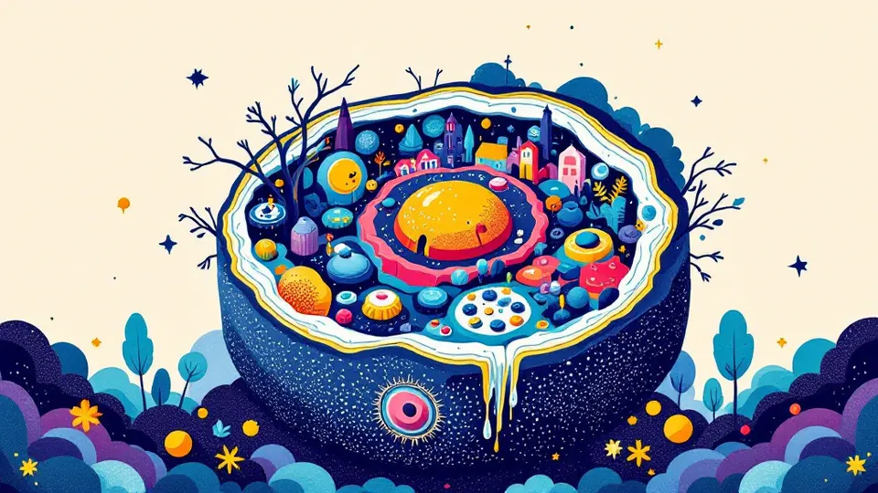
The flat, egg-shaped cell in your textbook is a simplified model. Real animal cells are three-dimensional. They come in many shapes, from long nerve cells to disc-shaped red blood cells, and they are packed with structures and dissolved chemicals. The diagram helps you learn the parts. Just remember that real cells are far more crowded and varied.
The main parts
These are the structures you need to know first:
- Nucleus: holds the cell's DNA, the instructions for making proteins. It controls the cell's activities by controlling which proteins are made.
- Cell membrane: a thin, flexible boundary that controls what enters and leaves the cell, such as glucose, oxygen and waste.
- Cytoplasm: the jelly-like fluid where many chemical reactions take place, helped by enzymes.
- Mitochondria: where aerobic respiration releases energy from glucose. Muscle cells contain many of them.
- Ribosomes: tiny structures where proteins are assembled. Some float freely, and others sit on a folded membrane system called the rough endoplasmic reticulum.
Many courses also mention the Golgi apparatus, which packages proteins for export. They may mention lysosomes too, which break down waste and worn-out parts. Check which ones your syllabus requires.
How the parts work together
No part works alone. The best way to understand a cell is to follow one job from start to finish.
Example: a cell in the pancreas making a digestive enzyme
1. The nucleus holds the gene for the enzyme. A copy of that instruction is sent out into the cytoplasm.
2. Ribosomes on the rough endoplasmic reticulum read the copy and build the enzyme, which is a protein.
3. The Golgi apparatus packages the enzyme into small membrane bubbles called vesicles.
4. The vesicles fuse with the cell membrane and release the enzyme outside the cell.
5. Throughout, mitochondria supply the energy for building and transport. The cytoplasm is where much of this movement happens.
Cause and effect: a cell that exports lots of protein needs many ribosomes and plenty of rough endoplasmic reticulum.
Animal cells versus plant cells
Plant cells have all the parts above, including mitochondria, because plants respire too. They also have three extras: a rigid cellulose cell wall, a large permanent vacuole filled with cell sap, and chloroplasts for photosynthesis. Animal cells have none of these. You will need chloroplasts in the next lesson.
What to do next time
When you meet a question about a cell part, give its function and link it to a process. For example, say what it does, then why the cell needs that done. When you see an unusual cell, ask which parts it would need lots of for its job. That habit turns memorised labels into real understanding.
Key takeaways
- The nucleus holds DNA and controls the cell by controlling which proteins are made.
- Mitochondria release energy from glucose, and ribosomes build proteins.
- Textbook cell diagrams are simplified models of crowded, three-dimensional cells.
Check your understanding
Written with AI help. Spotted a mistake? Tell us.